Hello Everyone, This will be my first blog but I hope you will like it
Disclaimer: This blog is just intended to be a guide and due to our individual differences, this may not guarantee for you to pass your subject, but this may serve as a guide and you may modify this according to your own preferences or study style. But hope it could help
Credits to PPCMD2 for teaching me how to use blogger for blog like this, to share some of his advice on how to create this blog
Dedicated to: Students from my alma mater, as I want to do my best to even a small blog can help to guide you.
As already stated in my previous blog, I already had mentioned the importance of visual learning for medical school but today, let us delve ourselves deeper as this blog will emphasize more on the importance of visual learning which will benefit you both from your practical exams and will help you studying for Long Exams, Revalida, and especally as long term preparation for Philippine Physician Licensure Exam.
**There will be no table of contents since this is just one whole topic but subtopics will be written in bold which will span to selected 1st year to 3rd year subjects
In general for active learning purposes, you may answer this questions what you have in mind this 5 general W and H questions
- What does a typical structure of a normal person or an disease entity look like?
- Where does this located?
- When will this appear?
- Who are the affected population?
- Why does this happened?
- How did this arrived at what do I see right now?
ANATOMY
For the Gross Anatomy part, it is important to VISUALIZE and IMAGINE the structures, imagination of the structures will not just help you at the laboratory for your practical exam in the Pictures in Online or Cadavers if Face to Face but also when taking the theoretical exam
Tip: while studying for the theoreticals, you may also start reviewing for the laboratory at the same time, this is for me as part of active learning for imagination at the same time I can save my time for the sake of other subjects
Ex,
In studying for the cranial nerves for the anatomy in terms of the foramen in terms of the skull, (ex. in the foramen ovale in the cranial base of the skull)
Taken and Modified from Abraham and McMinn Clinical Atlas of Anatomy 8th Ed.
I just posted it there since this picture resembles an actual practical exam which a pushpin or in that case a paper that is folded into the "Foramen ovale" but the question in reality is not just on answering the "Foramen ovale" but for example, What structures pass thru this structure? or What is the function of the structure that passes thru this structure?, see the sense is it is already a two way questions
Main question asked in the practicals: What is the structure of the function passing thru this ovale? (this is a one item question but in fact in reality its a two way question)
Two way questions is that
Initial question: what is the structure passes through that hole with the paper on it?
Stem or asked question: what is the structure of the function passing thru this hole?
To answer it we must go back to what we have learned in the lecture or theoretical aspect of the structures passing thru the foramen
What I did to have my imagination is that I will use an atlas (I use netter) besides the theoretical reading material so that I can imagine all the structures in that part
Taken from Netter Atlas of Human Anatomy 7th Ed.
Taken from Moore Clinically Oriented Anatomy 8th Ed.
As we can see from above, the table we can see of the structures which passes thru the Foramen ovale and as we all know the Mandibular nerve provides motor thru the muscles of mastications and provide sensory to the mandible. What I did here is I reviewed lecture and laboratory practicals at the same time
Also important in the everywhere but especially to the visceral organs also applied to skeletal organ is to know the relationship of an organ to a following organ structure
Ex. What is the structure that is superior to this and superior to that so know the relation like looking into a google map, except the google map is an atlas
For example, what is the structure that is superior to the first part of the duodenum
Taken from Netter Atlas of Anatomy 7th Ed.
Ok as you can see from above, is the duodenum in relation, look at the superior inferior laterial and medial view (take note it is important to learn your terms in anatomy) ex Supeior to the 1st part of duodenum is the liver, inferior to the 1st part is the head and neck of the pancreas lateral from the duodenum is the ascending colon and medially to the is the stomach and the abdominal aorta at the level of T12 and IVC
Cross Section of Abdomen at the level of T12 Vertebra
For looking at the anterior and posterior view, I would recommend if you cross or shift to the cross section, now look anterior to is the right colic flexure of the colon and posterior to it is the portal triad now if I will be given an exam relating to the structures and its relations now you can be given time to answer. I suggest you try it to other structures as well
Lastly for gross anatomy the most hard one (perhaps in my case is the tabulated OINA of muscles) but lately i found a way to simplify this, what I did is to compartmentzalize this muscles by looking at the atlas at the same time, since there is a common nerve, and common action that will supply these nerves
Lets take a look again at the arm compartment
Taken from Netter Clinically Oriented Anatomy 8th Ed.
Lets take the muscles for the arm for example again this is a tabulized version of the muscles involving the arm
Taken from Snell Clinical Anatomy by Regions 10th Ed.
By just looking at the table my comment that it is lengthy however visualizing the table would help in theoretical aspect
This is the compartmentalized version of the forearm, if studying OINA, the best way is to compartmentalize them
As you can see above there is the compartmentalized version you can see the compartment and how musculocutaneous nerve is drawn to them
But in the laboratory it will be different since it will be used by pushpins (in the actual face to face scenario in the practical exam) again correlate laboratory and lecture
Example you were asked about biceps the pushpin is in biceps and you will be asked for the nerve innervation, then you will refer to the table, however the practicals will not be like what you see in the atlas since this is just lecture (unless it is online)
But rather it will be like this
Taken from McMinn Clinical Atlas of Anatomy 8th Ed. pdf and see the difference of actual and a drawing
Next is the arteries and veins, the best way is to trace them and use atlas besides it, well its red and blue but in the practicals it will be different again, take note an artery will be lighter in color and veins will be darker in color
Taken from McMinn Clinical Anatomy 8th Edition
Take a look at this picture, in the reality it looks like a spaghetti without a sauce at all, in reality these are collection of nerves (in the Picture is the Brachial Plexus) and collection of arteries and veins supplying the upper thorax and the hand but to apply the determination
I just emphasized in this illustration the real color of an artery vs vein in real life but be careful but if it is solid like the no 3 without something side it is most probably a nerve (no, 3 is the superior trunk of the brachial plexus)
Speaking of the Brachial Plexus another active learning that i did is to draw the structure handwritten by maxing a diagram (you can search youtube videos for drawing these plexuses)
For Histology, now we will deal with the structures of the human body seen in microscope, again correlation of your lecture with the practicals in laboratory can also be part of active learning
For Example, lets take a look of the structure below
Taken from diFiore Atlas of Histology 13th
The best way is to
1. Know the organ
2. Know the function of the structure (can be correlated to other subject
correlate for example what is the function of the structure (ex. pointer is pointed the the glomerulus itself) then I can see its the glomerulus, i can see that there are red blood cells on the picture, I know from this subject and Physiology these filter blood from waste materials that is excreted renally now I have correlated the subjects, usually side questions can include many functions, use, disease that may be involve, so it is important to review alongside with lecture
Next is Embryology, in embryology as we can see in both textbooks and notes, that it is written in a narrative format like narrator telling a story to the audience because its basically story telling, but not fiction but a story of a human or an embryo or fetus is formed.
Take a look at this
Taken from Moore The Developing Human 10th Ed.
As you can see on that picture it is a series of story and the narration is found on the text itself, how the stomach form from a simple tube to become the stomach we saw as adults and the narrative is down below, in embryology illustration and imagination is important the goal here is not just imagination but making it what I call a mind video meaning like a video playing on how it can form and applying it into the lab also this story, if someting is wrong I can apply it in Pediatrics, and Pediatric Surgery subject
Typical Laboratory slides seen in Medical Schools for Embryology
For the laboratory apply ex fate of that tube or gut tube is the stomach apply what you had learned in the laboratory
PHYSIOLOGY
Physiology is understanding again, that its textbooks are writen in a narrative format like a story also, the best trick that works for me is to read the book with looking at the illustration afterwhich check the illustration again and explain it or simplify in the way that I could understand or this is one of the subjects for me that I can use Tagalog or mother tongue to understand this one
Taken from Berne and Levy Physiology 7th Ed.
In the standard textbook, this will be a very long narrative text but you can create both a story shortened and in your own mother tongue.
Simplify like "if Na is outside K is inside then Na goes in papasok sa loob since mas maraming Na sa labas sa passive if inside to outside then active diffusion pipilit lumabas so dapat me magtutulak which is si ATP gagawa nian" now i simplified that picture above, same can be done with these graphs
Now lets deal with some complex ones
Taken from Guyton and Hall Medical Physiology 14th Ed.
You can say, too many lines too many things to remeber but priming or bisecting this graph will be very helpful have a anatomy of the heart and try to understand it and translate in the mother tongue or Tagalog like this:
"Unahin natin sa systole, sa sysole as u can see lets say ung red sa taas is pressure or puwersa of blood while blue sa baba is the volume or dami ng blood both sa VENTRICLE since ung heart is mase up of muscles involuntariy automiatic from SA node pipiga ung heart mo so ang nanyayari nagkakaroon ng pressure o puwersa pero naiipon at naiipon pa yan so un ang ISOVOLUMIC CONTRACTION pero volume sill ganun parin tho on the other side sa atrium mo unti unti ng naglalagay ng dugo jan kayabtignan mo ung curve tumataas siya forming the C wave pero once na sobrang lakas na ng pressure bubukas ang aortic valve mo sa (ecg ang pagpiga ng ventricle is qrs complex) hayun pressure goes up punta sa ascending aorta ito na ang EJECTION mo ung dugo kaya ung volume tignan nio ung blue bumababa until bumaba ang dugo bagsak ang aortic pressure then tignan mo atria mo on the broken line nagiipon ipon yan ng dugo from pulmonary vein w/c carries oxygenated blood kaya pataas siya siempre pababa na rin ang daloy ng kuryente sa ventricle kaya nagnenegative T wave or repolarization nanyayari, going back sa dugo once bumaba ang pressure ng dugo mula ventricle aortic valve closes forming the incisura pero on tje other hand di pa ganong kalakas ang pressure ng dugo sa atrium kaya sa ISOVOLUMIC RELAXATION bagsak sion then magiipon ipon yan pag malakas tjen pipga si atrium (p wave sa ecg) taas atrial pressure bukas ang mitral valve or av valve nio rapid inflow of blood tignan nio ang volume tumataas ang dugo aa ventricle kaya pataas ang kulay blue state of diastasis then systole again forming another cycle"
**Credits and Rewritten with Permission from PPCMD2
Here what I did is 3 things: correlate, simplify, and explained in mother tongue now let me give you assignment or exercises, can you do this 2 in your physiology how about this two problems for practice, try to correleate simplify and explain in mother tongue or Tagalog
For the simple one
For a complex one
BIOCHEMISTRY
This subject deals a lot of cycles and structure ok important here are the substrates, enzymes, inhibitors, products, especially the important ones the finished product ansewring the questions
1. What is the product and the use of the product
2. How does it is produced
3. What will stop the production or decrease of the quality of the product
4. Which step is irreversible, energy producing and energy consuming product
5. How important is the products produced to the human body
Example of these are how to synthesize proteins carbs fats, and how to use them into energy or storage for the human body, this is for me where your background in basic Economics will come in since rules in economics apply in the biochemical reactions of the human body
Fatty Acid Synthesis. Taken from Lehninger Biochemistry 8th Ed
Beta Oxidation, Harper's Illustrated Biochemistry 32nd Ed.
Now the best way approach to this guys is to read the text along while looking this cycles as this will help you visualize cycle and focus the most important ones (same goes in the cycle). The most important ones in the cycle will include
1. Irreversible or Rate-limiting reactions
2. Substrates or Co-substrates in a rate limiting reactions
3. Phase of the Pathway
4. no or ATPs, NAD and FAD oxidized or consumed, to compute energy
5. Inhibitors or Drugs/Xenobiotics that Inhibit Rate limiting step
6. Important diseases if something happens in that pathway
For structures, the best way is to visualize the structures and what composed it like in the Proteins of Primary, Secondary, Tertiary, and Quarternary Structures. or DNA how its composed of
Taken from First Aid USMLE Step 1
Also mnemonics will also help, visiting USMLE Step 1 First aid can also help for your mnemonics which can be helpful in your long exams
CLINICAL MEDICINE I OR PHYSICAL DIAGNOSIS
This is a subject where what you had learned in your first year subjects will be applied already to actual patients, you will learn how to approach patients, ideally this is learned thru experience for active learning but important to visualize here is the structures that involve for example in the lungs
Lung percussion, taken from Bates Guide to Physical Examination 13th Ed.
This subject shall involve your 5 senses in terms of applying your subjects not limited only to visualization (unless teleconsultation) apply it
Here's another example
Nail Plate Disorders, taken from DeGowin Diagnostic Examination 9th Ed,
Heart Murmurs, taken from DeGowin Diagnostic Examination 9th Ed.
This are the things that the key to learn is during 4th year or internship since actually having cases like this will train you and is a form of active learning tho youtube can help as supplement, but experience nothing can beat that
PATHOLOGY
A typical Philippine medical school has 3 (or 4) pathology in some General, Systemic, Clinical (and Surgical) in some but usual scenarios here is that it is Physiology, Biochem in reverse of the normal is abnormal important is to visualize it by using an atlas, or same with physio, practice using mother tongue to simplify it tho what cannot be simplified are memorizable facts and pathgonomic hallmarks
Mechanisms of Glomerular injury taken from Robbins Cotran Pathologic Basis 10th
Applying General Pathology, Physiology, and Immunology and Microbiology, you can explain what in this structure by using the picture above leading to this
PSGN. Taken from Robbins 10th Ed.
Here is the example of defaced glomerulus as compared to above, as you can see it is defaced, full of lymphyocytes, immunofloresence shows Immunoglobulins attached to the glomerulus itself apply in your Clinical Medicine, this will present as hematuria or blod in the urine since filtration system is damaged so visualize with atlas (Robbins has its own atlas also). For the practical exam it is important also to review lecture also, important in pathology is the characteristic morphology, genes if applicable
Same goes to Clinical Pathology where visualizations will be like first years but with application onto it not just visualize but memorize very very important facts in there
Protein Determination via SDS-PAGE, Taken from Henry's Clinical Diagnosis 24th Ed.
Goal there is apply the principle of SDS-PAGE via the Electrophoresis or Charges present in the ion of each biomolecule (refer to Basic Chemistry and Biochemistry for good background also). and for Hematology correlate 1st year with both of clinical pathology. I may be late to tried and prove this but the 3 rules of Physiology, Correlate, Simplify, and tagalog can also be applied in this subject (but do not attempt this on CPCs if you're gonna do this, only on reviews)
MICROBIOLOGY
Microbiology is also a visualization subject aside from the knowing the structure you have to know the respective culture media for them respectively, tabulating them can also be good since it helps for the student to compare the different and what is important in that micro-organism such as the disease, specific test
'For practical exam it is also important to know the lecture and the culture media or microscopic feature of the specimen as like in Anatomy since side question can also be asked like disease, agar that can be used specific toxin etc.

Algorithms for bacteria
Algorithm for Viruses
(For the laboratory for those who wish to create a transes/reviewer for practicals I would suggest that you take a picture or if online you can use these atlases for reference such as De la Maza Color Atlas and Photographic Atlas of Microbiology

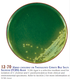
Sample Microscopic and Culture for Vibrio Taken from Leboffe Pierce Photographic Microbio 4th Ed
Like Anatomy and Pathology, I would like to recommend to use Atlas alongside Microbiology so that it is a two way review for both lec and lab
If you are a fan of comic strip you can do this style
Taken from Clinical Microbiology made Ridiculously Simple 9th Ed.
Taken from Clinical Microbiology made Ridiculously Simple 9th Ed.
PARASITOLOGY
For Parasitology, it is the same principle such as now it involves lifecycle, and knowing some important anatomic parts of the parasite, aside from the mother text, using atlas can also be advisable to aside from supplementing your learning you can also use this for either answering your lab manuals, creating transes for both practicals and great aid for lecture
Ex. Giardia Lamblia parasite
Taken from CDC Lifecycle of Giardia Lamblia
To further aid your lectures and allow you to study for practicals at the same time, using atlas at the same time can help
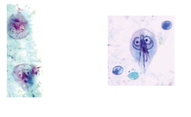
Cyst and Trophozoite forms drawings of G. lamblia, from Medical Parasitology Self Insttructional TExt
Cyst and Trophozoite forms of G. lamblia, from Gacia, Practical Guide to Diagnostic Parasitology
DERMATOLOGY
In Dermatology, it is most recommended for me in the med school as studying dermatology subject to look at the skin lesions since they almost look like the same and look for the characteristic lesion
Ex. in Acne vulgaris Closed comedones appear as cream to white, slightly elevated, small papules and do not have a clinically visible orifice, to better understand this we need atlas and picture from derma books
Acne lesions taken from Fitzpatrick Dermatology
NEUROLOGY
in the Neurology part the way that may work is to know your Neuroanatomy and the Homoculus since this will give you edge to the lesions that you and signs and symptoms of patient, a pathology background will also be good and correlate it with clinical medicine
Spinothalamic pathway for Sensory System from Snell Anatomy 8th Ed.
CLINICAL MEDICINE II/ PEDIATRICS/ SURGERY/OB-GYNE
For the Clinicals, the way that may work are those tables and clinical pathways found in CPGs and textbooks which will make the topics easier, off course still reading text, analyzing and memorizing most important parts only will be a good offset. Remembering the pathological gross and microscopic appearance can also help you alongside with good Pharmacology background
Examples of some
Classification of UC from HPIM 20th Ed.
Amenorrhea algorithm
For the Surgery the way that I did is to know the location of the disease and look at the pictures on the book either Schwartz or Sabiston to map of where the diseased entity and for it to be resected does the case presentation will be done. Example is colectomy for adenocarcinomas
The way that I use is I did record the lecture, also I jot down notes handwritten so that I can plan ahead on what to review on the transes or if I have time, textbook itself, also I found out that it has advantage in terms of I have a short time or short breaks already since I can use the lecture itself as like 1st reading and the after lecture reading itself is the 2nd reading to avoid having backlogs, since handwriting notes is also aside from planning on how to review the subject, it is also a form of active learning for the retention of information or knowledge which I will need in the exam.
RADIOLOGY
Radiology has its own practical exam also, the way I remembered that it was it that to read the text while looking at the plates or pictures of the plate being used and apply what I had learned from other subjects to derive from that look in the Radio for reading. Also apply what you had learned in the Gross Anatomy since it will help a lot in terms of correlating the disease to the location
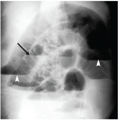
Ex. for practicals a patient comes in with epigastric abdominal pain associated with difficulty of defecation PMHx: patient had undergone ileostomy secondary to a tumor upon auscultation it is 35/min bowel movement RLQ and 14/min LLQ and Abdominal X-ray shows above, air fluid level with proximal ends dilated signifying SBO. now i reviewed Surgery, Radiology and pracs at the same time
OPHTHALMOLOGY
For Ophthalmology usually is application of other subject, but the visualization for eye is also important since eye lesions as my experience is look like the same then looked without history, eye visualization can be helpful. While doing with transes, I would recommend using Kanski's Clinical Ophthalmology pictures since it also being an atlas of its own about on how to approach eye disorders, and can be used to supplement the lectures which is Vaughn General Ophthalmology
Ex.
Bacterial vs Fungal Keratitis taken from Kanski's Clinical Ophthalmology 9th Ed.
LEGAL MEDICINE/MEDICAL JURISPRUDENCE
In Medical Jurisprudence both the green book (used in medical schools) and Solis (used in the PLE) uses analytical type of statements in their textbooks, the way that I did is to analyze the scenario in relation to medicine and apply the laws and ethics, doing tables can be helpful also, look at the examples it can also be helpful
In Legal Medicine, there is a lot of tables and illustrations, regarding like example the difference of entrance and exit wounds, table of alcohol percentage of drunk driving, fingerprints etc. what I did was
to visualize them and try to rewrite some complex tables handwritten to the index card
Sample Incised wound taken from Solis Legal Medicine
Sample table from Solis Legal Medicine
Take note of how this table can be rewritten in an index card for better memory
Final Message: correlation of one subject with visualization plus active learning as per experience will be a good way for a better retention


Mga Komento
Mag-post ng isang Komento